+3901118837572
Via Amedeo Gioliti,5 Pianezza (TO) ITALY
+3901118837572
Via Amedeo Gioliti,5 Pianezza (TO) ITALY
Ultrasonography is a type of medical imaging that uses high-frequency sound (ultrasound) waves to produce images of internal organs and other tissues.
During an ultrasound, a device called a transducer converts electrical current into sound waves, which are sent into the body’s tissues. Sound waves bounce off structures in the body and are reflected back to the transducer, which converts the waves into electrical signals. A computer converts the pattern of electrical signals into an image, which is displayed on a monitor and recorded as a digital computer image. No x-rays are used, so there is no radiation exposure during ultrasonography.
Procedure for Ultrasonography
If certain parts of the abdomen are being examined during an ultrasound, people may be asked to refrain from eating and drinking for several hours before the test. Normal gas build up in the intestines from eating or drinking can sometimes make ultrasound images difficult to obtain. In other instances, such as an ultrasound of female reproductive organs, women may be asked to drink a large amount of fluid to fill their bladder.
Usually, the examiner places thick gel on the skin over the area to be examined to ensure good sound transmission. A handheld transducer is placed on the skin and moved over the area to be evaluated. Uses of Ultrasonography
Ultrasound images are acquired rapidly enough to show the motion of organs and structures in the body in real time (as in a movie). For example, the motion of the beating heart can be seen, even in a fetus.
Ultrasonography is effectively used to check for growths and foreign objects that are close to the body’s surface, such as those in the thyroid gland, breasts, testes, and limbs, as well as some lymph nodes.
Ultrasonography is effectively used to image internal organs in the abdomen, pelvis, and chest. However, because sound waves are blocked by gas (for example, in the lungs or intestine) and by bone, ultrasonography of internal organs requires special skills. People who have been specifically trained to do ultrasound examinations are called sonographers.
Ultrasonography is commonly used to evaluate the following:
Heart: For example, to detect abnormalities in the way the heart beats, structural abnormalities such as defective heart valves, and abnormal enlargement of the heart’s chambers or walls (ultrasonography of the heart is called echocardiography)
Blood vessels: For example, to detect dilated and narrowed blood vessels
Gallbladder and biliary tract: For example, to detect gallstones and blockages in the bile ducts
Liver, spleen, and pancreas: For example, to detect tumors and other disorders
Urinary tract: For example, to distinguish benign cysts from solid masses (which may be cancer) in the kidneys or to detect blockages such as stones or other structural abnormalities in the kidneys, ureters, or bladder
Female reproductive organs: For example, to detect tumors and inflammation in the ovaries, fallopian tubes, or uterus
Pregnancy: For example, to evaluate the growth and development of the fetus and to detect abnormalities of the placenta (such as a misplaced placenta, called placenta previa)
Musculoskeletal structures: For example, to detect swelling within a joint or a torn ligament
Ultrasonography can also be used to guide doctors when they remove a sample of tissue for a biopsy. Ultrasonography can show the position of the biopsy instrument, as well as the area to be biopsied (such as a mass). Thus, doctors can see where to insert the instrument and can guide it directly to its target.
Doppler ultrasonography
Doppler ultrasonography uses changes that occur in the frequency of sound waves when they are reflected from a moving object (called the Doppler effect). In medical imaging, the moving objects are red blood cells in the blood. Thus, Doppler ultrasonography can be used to evaluate
-Whether blood is flowing through blood vessels
-How fast it flows
-Which direction it flows in
Doppler ultrasonography is used :
-To evaluate how well the heart is functioning (as part of echocardiography).
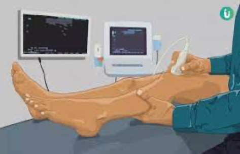
-To detect blocked blood vessels, especially in leg veins, as in deep vein thrombosis, when veins are blocked by a blood clot
-To detect narrowed arteries, especially the carotid arteries in the neck, which carry blood to the brain
Spectral Doppler ultrasonography
Spectral Doppler ultrasonography shows blood flow information as a graph. It can be used to assess how much of a blood vessel is blocked.
Duplex Doppler ultrasonography
Duplex Doppler ultrasonography combines spectral and B-mode ultrasonography.
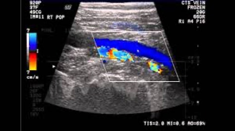
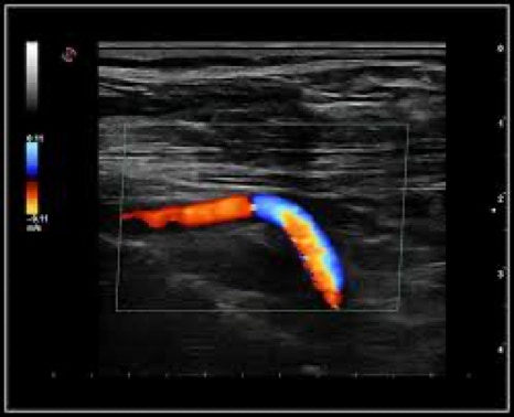
Color Doppler ultrasonography
For color Doppler ultrasonography, color is superimposed on the shades-of-gray image of blood flow produced by Doppler ultrasonography. The color indicates direction of blood flow. Red may be used to indicate flow toward the transducer, and blue may be used to indicate flow away from the transducer. The brightness of the color indicates how fast the blood is flowing.
Color Doppler ultrasonography can help assess the risk of stroke because it helps doctors identify and evaluate narrowing or blockage of arteries in the neck and head. The procedure is useful for evaluating people who have had a transient ischemic attack or stroke and people who have risk factors for atherosclerosis but no symptoms. Color Doppler ultrasonography is also used to assess blood flow to internal organs and tumors.
Questo sito web fa uso di cookies. Si prega di consultare la nostra informativa sulla privacy per i dettagli.
Non consentire
OK
